Ectopic adenoma of parathyroid gland
Payman Maleki, Sokolina I.A., Pasha S.P.
Moscow Medical Academy, Moscow, Russia Pogodinskaya Str, 1, E-mail: pmarad@yahoo.com
–Я–∞—Ж–Є–µ–љ—В
–Т–Њ–Ј—А–∞—Б—В: 55 years
–Я–Њ–ї: –ґ–µ–љ—Б–Ї–Є–є
–Р–љ–∞–Љ–љ–µ–Ј
Presenting to you is a 55-year old female with chief complaint of general weakness and easy fatigability accompanying with headache, morning nausea, significant sweating, mouth and skin dryness, abdominal protrusion, episodes of liquid bloody stool, decreased visual acuity and incomplete bladder emptying.
In patientТs past history thyroid gland changes were found during sonography in 2000 but she didnТt receive any treatment from 2001 to 2002. It should be mentioned that referred recent complaints appeared after menopause. Meanwhile she developed multi glandular goiter in 2004 so received 200 mg of Iodine during 6 months. The level of TSH (Thyroid Stimulating Hormone) and free T4 became normal in 2005.
During sonography of Thyroid gland sings of chronic autoimmune thyroiditis and adenoma of right upper
parathyroid gland were detected so recommended to perform Scintigraphy and CT which will be discussed
in following paragraphs.
–Т—Л–њ–Њ–ї–љ–µ–љ–љ—Л–µ –Є—Б—Б–ї–µ–і–Њ–≤–∞–љ–Є—П
Densitometry, scintigraphy of parathyroid gland, spiral CT (HiSpeed CT/I General Electric,collimation: 3 mm., table feeding: 3 mm, contrast media: Omnipak 350 IU/ml, injection rate 3 ml/s).
–Ю–±—Б—Г–ґ–і–µ–љ–Є–µ
Densitometry: initial osteopenia of left femur. Scintigraphy of parathyroid gland:
neoplasm (adenoma) in right superior parathyroid gland. Signs of ectopic parathyroid adenoma in mediastinum. Spiral CT: in the series of axial tomography of chest at the middle of anterior mediastinum, 3.6 cm below the upper level of manubrium, a soft tissue well defined ovoid mass (50 u.H.) was appeared with the size of 14 x 12 x 9 mm (fig. 2a).
This mass is differentiated from arteries around by lower enhancement after injection of contrast media (fig. 2b, c).
–Ю–Ї–Њ–љ—З–∞—В–µ–ї—М–љ—Л–є –і–Є–∞–≥–љ–Њ–Ј
Ectopic adenoma of parathyroid gland
–Р–і—А–µ—Б —Б–ї—Г—З–∞—П –≤ –Є–љ—В–µ—А–љ–µ—В–µ
http://www.tomography.ru/cases.php?id=41
|
–†–Є—Б—Г–љ–Њ–Ї 1a 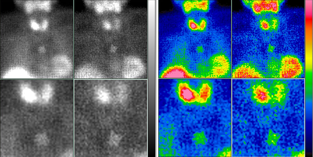
–†–Є—Б—Г–љ–Њ–Ї 2a 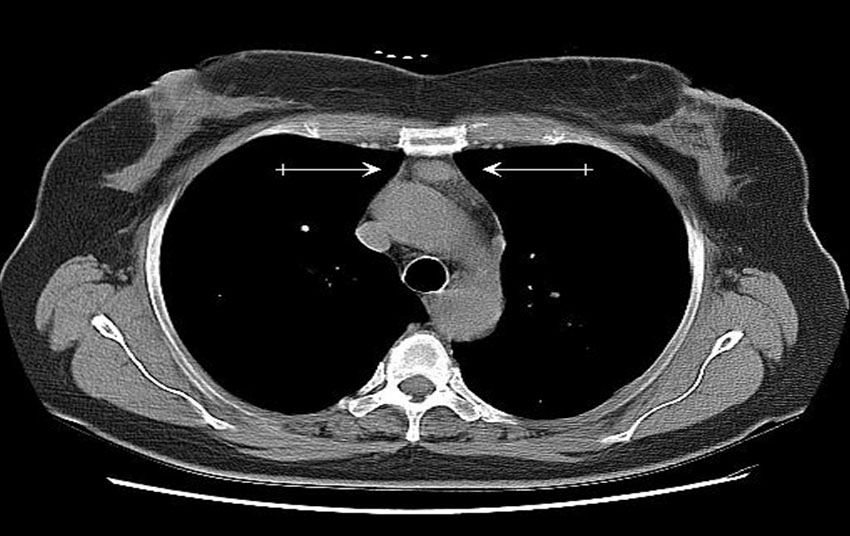
–†–Є—Б—Г–љ–Њ–Ї 2b 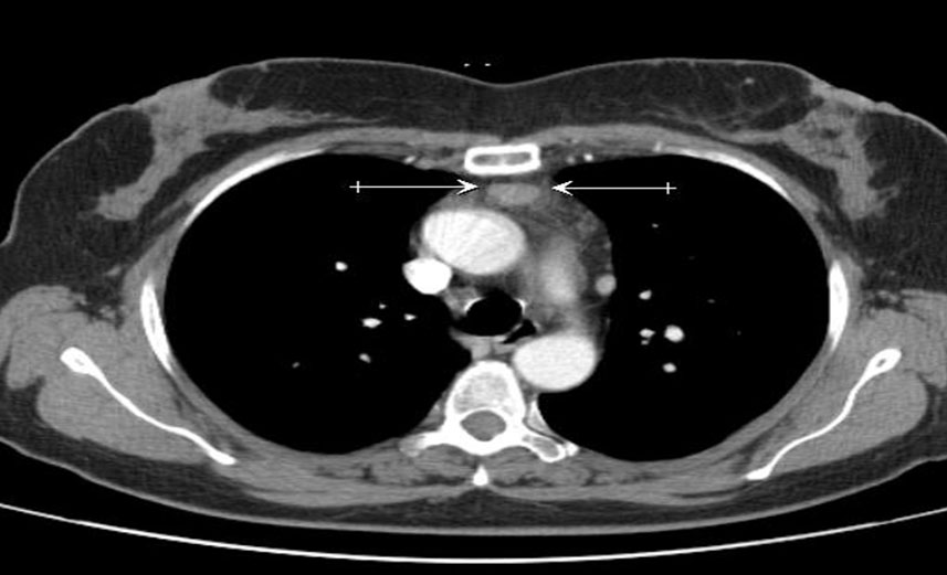
–†–Є—Б—Г–љ–Њ–Ї 2c 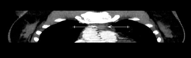
|